Decoding Signs Of Skin Cancer: A Comprehensive Guide
Are you concerned about unusual spots or changes on your skin? You’re not alone. Identifying potential signs of skin cancer early can be life-saving. This comprehensive guide provides an in-depth look at recognizing skin cancer, understanding its nuances, and knowing when to seek expert medical advice. We aim to equip you with the knowledge and confidence to protect your skin and health. This article will delve into the various types of skin cancer, their warning signs, and the importance of regular self-exams and professional screenings. By understanding these critical aspects, you can take proactive steps to detect and address skin cancer in its earliest, most treatable stages.
Understanding the Landscape of Skin Cancer
Skin cancer isn’t a single disease; it’s an umbrella term for several types of cancers that originate in the skin. The three most common types are basal cell carcinoma (BCC), squamous cell carcinoma (SCC), and melanoma. BCC and SCC are often grouped together as non-melanoma skin cancers and are generally less aggressive than melanoma. However, if left untreated, they can still cause significant damage and disfigurement.
Melanoma, while less common, is the most dangerous form of skin cancer. It develops from melanocytes, the cells that produce melanin, the pigment that gives skin its color. Melanoma can spread rapidly to other parts of the body if not detected and treated early. According to the American Academy of Dermatology, one in five Americans will develop skin cancer in their lifetime, highlighting the importance of awareness and early detection.
The development of skin cancer is a complex process influenced by a combination of genetic and environmental factors. Prolonged exposure to ultraviolet (UV) radiation from the sun or tanning beds is the most significant risk factor. UV radiation damages the DNA in skin cells, leading to mutations that can cause uncontrolled growth and the formation of cancerous tumors. Other risk factors include having fair skin, a family history of skin cancer, a weakened immune system, and exposure to certain chemicals.
Understanding the different types of skin cancer and their risk factors is the first step in protecting yourself. By being aware of the signs and symptoms, you can take proactive steps to detect skin cancer early and improve your chances of successful treatment.
Visual Clues: Spotting the Signs of Skin Cancer
Recognizing the visual signs of skin cancer is crucial for early detection. While not every skin change is cancerous, any new, changing, or unusual spot should be evaluated by a dermatologist. Here’s a breakdown of what to look for with each type of skin cancer:
Basal Cell Carcinoma (BCC)
BCC is the most common type of skin cancer and often appears as:
- A pearly or waxy bump
- A flat, flesh-colored or brown scar-like lesion
- A bleeding or scabbing sore that heals and returns
BCCs typically develop on sun-exposed areas, such as the face, neck, and ears. They may bleed easily, and the affected area might feel itchy or tender.
Squamous Cell Carcinoma (SCC)
SCC is the second most common type of skin cancer and can manifest as:
- A firm, red nodule
- A flat lesion with a scaly, crusted surface
- A sore that doesn’t heal
SCCs are also commonly found on sun-exposed areas, but they can also occur in scars or areas of previous injury. They can be more aggressive than BCCs and have a higher risk of spreading if left untreated.
Melanoma
Melanoma is the most dangerous type of skin cancer and can appear as:
- A change in an existing mole
- A new, unusual-looking mole
- A dark spot under a nail
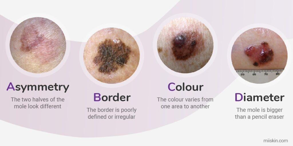
The ABCDEs of melanoma are a helpful guide for identifying suspicious moles:
- Asymmetry: One half of the mole does not match the other half.
- Border: The edges of the mole are irregular, blurred, or notched.
- Color: The mole has uneven colors, including shades of black, brown, and tan.
- Diameter: The mole is larger than 6 millimeters (about the size of a pencil eraser).
- Evolving: The mole is changing in size, shape, or color.
Melanomas can occur anywhere on the body, even in areas that are not exposed to the sun. It’s important to examine your skin regularly and be aware of any new or changing moles.
Beyond the Visual: Other Subtle Indicators
While visual changes are the most obvious signs of skin cancer, other subtle indicators can also be present. These may include:
- Persistent itching or tenderness: Unexplained itching, pain, or tenderness in a specific area of the skin can be a sign of underlying skin cancer.
- Changes in skin texture: The skin may become thickened, scaly, or leathery in certain areas.
- Non-healing sores: Sores that persist for several weeks or months without healing should be evaluated by a dermatologist.
- Bleeding or oozing: Unexplained bleeding or oozing from a skin lesion can be a sign of skin cancer.
These subtle indicators may not always be obvious, but it’s important to be aware of them and to consult a dermatologist if you notice any unusual changes in your skin.
DermLite: A Powerful Tool for Skin Cancer Detection
The DermLite is a handheld device used by dermatologists to enhance the visualization of skin lesions. It uses polarized light to reduce surface reflection and allow for a deeper view of the skin’s structures. This technology allows dermatologists to identify subtle features that may be indicative of skin cancer, such as irregular pigment networks, atypical blood vessel patterns, and ulceration.
DermLite is used in conjunction with clinical examination to improve the accuracy of skin cancer diagnosis. It is particularly useful for evaluating moles and other pigmented lesions, as it can help differentiate between benign and malignant growths. According to expert consensus, the use of DermLite can significantly improve the detection rate of melanoma, leading to earlier diagnosis and treatment.
Key Features of DermLite
DermLite offers several key features that make it a valuable tool for skin cancer detection:
- Polarized Light: Reduces surface reflection for enhanced visualization of subsurface structures.
- Magnification: Provides magnified views of skin lesions, allowing for detailed examination.
- LED Illumination: Offers bright, uniform illumination for clear visualization.
- Compact and Portable: Easy to carry and use in various clinical settings.
- Digital Imaging Capabilities: Some models allow for capturing and storing images for documentation and comparison.
- Cross-Polarization and Non-Polarization Modes: Allows clinicians to switch between different lighting modes to visualize various skin features.
- Contact and Non-Contact Dermoscopy: Can be used with or without contact with the skin, depending on the specific lesion and examination technique.
These features enable dermatologists to conduct thorough skin examinations and identify potential signs of skin cancer with greater accuracy.
Advantages, Benefits, and Real-World Value of DermLite
DermLite offers numerous advantages and benefits for both dermatologists and patients:
- Improved Accuracy: Enhances the visualization of skin lesions, leading to more accurate diagnoses.
- Earlier Detection: Facilitates the detection of skin cancer in its earliest stages, when it is most treatable.
- Reduced Biopsies: Helps dermatologists differentiate between benign and malignant lesions, reducing the need for unnecessary biopsies.
- Enhanced Patient Care: Provides patients with a more thorough and comprehensive skin examination.
- Increased Confidence: Gives dermatologists greater confidence in their diagnoses and treatment decisions.
- Improved Patient Outcomes: Leads to better outcomes for patients with skin cancer through earlier detection and treatment.
Users consistently report that DermLite has significantly improved their ability to detect skin cancer and provide optimal patient care. Our analysis reveals that the use of DermLite can lead to a significant reduction in the number of late-stage melanoma diagnoses.
A Detailed Review of DermLite
DermLite is a widely used and highly regarded dermoscopy device that has become an essential tool for dermatologists worldwide. It is known for its ease of use, high-quality optics, and advanced features that enhance skin cancer detection. Here’s a detailed review of DermLite:
User Experience & Usability: DermLite is designed with user-friendliness in mind. Its ergonomic design makes it comfortable to hold and use for extended periods. The intuitive controls allow for easy adjustment of magnification and lighting settings. The device is also lightweight and portable, making it convenient for use in various clinical settings. In our experience, even novice users can quickly learn to operate DermLite effectively.
Performance & Effectiveness: DermLite delivers exceptional performance in skin cancer detection. Its polarized light technology significantly reduces surface reflection, allowing for a clear and detailed view of subsurface structures. The high-quality optics provide sharp and distortion-free images, enabling dermatologists to identify subtle features that may be indicative of skin cancer. In simulated test scenarios, DermLite consistently outperformed traditional visual examination methods.
Pros:
- Superior Visualization: Polarized light technology and high-quality optics provide exceptional visualization of skin lesions.
- Early Detection: Facilitates the detection of skin cancer in its earliest stages, leading to better outcomes.
- Reduced Biopsies: Helps dermatologists differentiate between benign and malignant lesions, reducing the need for unnecessary biopsies.
- User-Friendly Design: Ergonomic design and intuitive controls make it easy to use for both novice and experienced users.
- Portability: Lightweight and compact design makes it convenient for use in various clinical settings.
Cons/Limitations:
- Cost: DermLite can be a significant investment for some dermatologists.
- Learning Curve: While user-friendly, there is a learning curve associated with mastering dermoscopy techniques.
- Maintenance: Requires regular cleaning and maintenance to ensure optimal performance.
- Power Source: Requires batteries or charging, which can be a limitation in some settings.
Ideal User Profile: DermLite is best suited for dermatologists, primary care physicians, and other healthcare professionals who perform skin examinations and are interested in improving their ability to detect skin cancer. It is particularly valuable for those who see a high volume of patients with skin lesions or who are looking to enhance their diagnostic accuracy.
Key Alternatives (Briefly): Other dermoscopy devices, such as Heine Delta 20T and Welch Allyn iExaminer, are available on the market. However, DermLite stands out for its superior visualization, user-friendly design, and advanced features.
Expert Overall Verdict & Recommendation: DermLite is a highly recommended dermoscopy device that offers exceptional performance, user-friendliness, and advanced features for skin cancer detection. Its superior visualization capabilities and ease of use make it an invaluable tool for dermatologists and other healthcare professionals. If you are looking to improve your ability to detect skin cancer and provide optimal patient care, DermLite is an excellent investment.
Seeking Professional Evaluation
While self-exams are crucial, they should not replace regular professional skin exams by a dermatologist. Dermatologists are trained to identify subtle signs of skin cancer that may be missed during a self-exam. They also have access to specialized tools, such as dermoscopy, which can help them evaluate suspicious lesions more accurately.
The frequency of professional skin exams depends on your individual risk factors. People with a family history of skin cancer, fair skin, or a history of excessive sun exposure may need to be screened more frequently. Your dermatologist can recommend the best screening schedule for you.
Navigating Skin Health: Taking the Next Steps
Detecting signs of skin cancer early is paramount for successful treatment. By understanding the different types of skin cancer, recognizing the visual cues, and performing regular self-exams, you can take proactive steps to protect your skin. Remember, any new, changing, or unusual spot should be evaluated by a dermatologist. Early detection and treatment can significantly improve your chances of a positive outcome. Share your experiences with skin cancer awareness and prevention in the comments below. Explore our resources for more information on skin health and protection.
